Radiology for Dummies
Understanding different healthcare imaging types
We’re not calling you a dummy. Understanding the different types (called “modalities” in the medical world) of imaging done by radiology techs is not an easy task. But at Singing River Health System, we believe the more you understand about your health and the resources available to you, the more you can play an active role in your healthcare.
Let’s break down some of the most common radiology procedures in a way we can all understand them.
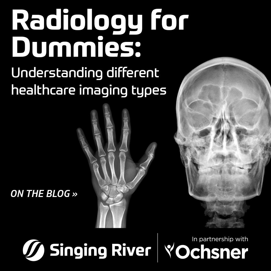
X-Ray
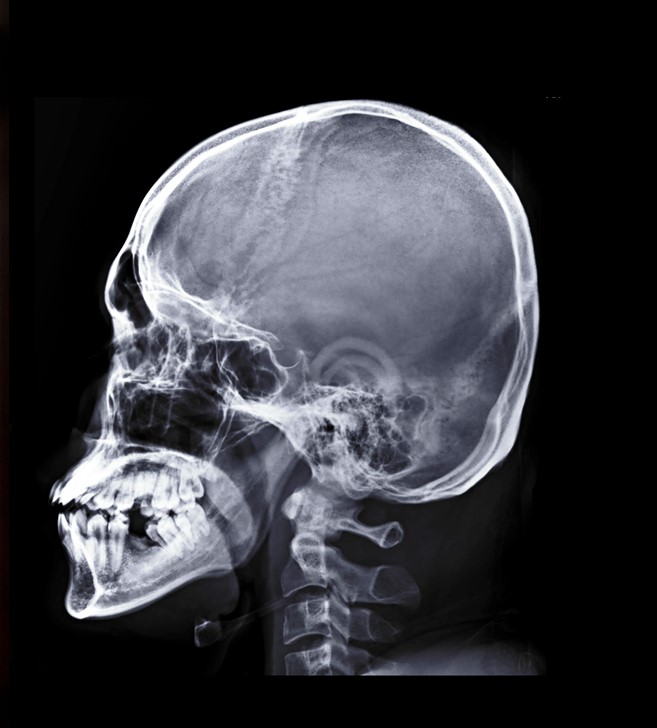
What is an X-Ray?
An X-Ray, also called a radiograph, captures images in black and white by sending radiation through the body. More dense areas and areas containing a lot of calcium block the radiation, making those areas appear white on the image. Softer tissues allow varying amounts of the radiation through and appear gray-black on the images.
What are X-Rays used for?
X-Rays are ideal for spotting issues in bones, such as fractures, dislocated joints, alignment issues, and narrowed spaces within joints. Subtle bone injuries like inflammation, tendon or ligament tears, or other soft-tissue issues are generally not apparent via X-Ray, but your provider may order an X-Ray to rule out a fracture.
CT Scan
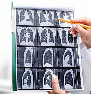
What is a CT Scan?
A CT Scan, or computer tomography scan, is similar to an X-Ray in that it captures images by sending radiation through the body. However, CT Scans offer much more detail, capturing “slices” of X-Ray type images to create 3D, 360-degree images of the body’s structures.
“If you’re looking at a loaf of bread, and the slices, that’s what a CT does. It breaks your body down, instead of looking at a 2 dimensional [image], you’re looking at a 3 dimensional loaf of bread.”
Jarrod Fetters, Singing River Radiology Manager
What are CT Scans used for?
CT Scans are ideal for looking at bony and muscular structures, as well as internal organs. Since they take only one minute to perform, they are ideal for emergency situations. Oftentimes, CT Scans are used to spot blood clots, subtle bone fractures not visible via X-Ray, injuries to organs, abscesses, and small lung nodules.
MRI
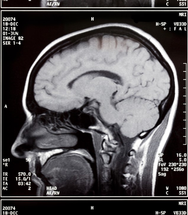
What is an MRI?
An MRI, or magnetic resonance imaging, does not use radiation. Instead, it uses a large, powerful magnet to pass radio waves through your body. Essentially, water molecules in your body react to the magnetic field to create signals that are turned into images. Like CT Scans, MRIs produce “slices” of images that can be combined to create 3D images of the body’s structures, however, given the different method, it can capture different things.
What are MRIs used for?
MRIs are useful for providing highly detailed images of a variety of the body’s structures, including muscular structure, soft tissues, nerves, and blood vessels. MRIs can spot issues that are too subtle for an X-Ray and can help identify cartilage loss, inflammation, nerve injuries, stroke, tumors, torn ligaments or tendons, and other abnormalities or functions of the internal organs. MRIs are very good at exposing slight differences between types of tissue that may not be evident via X-Ray or CT Scan.
Ultrasound
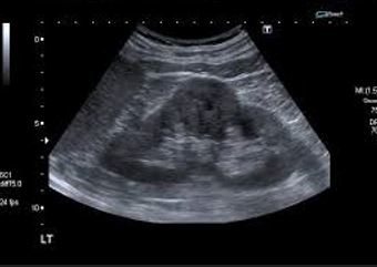
What is an Ultrasound?
An Ultrasound is an imaging method that uses sound waves to create images of structures inside your body.
What are Ultrasounds used for?
Most commonly, you might think of the static-y images of babies printed out and pinned to the fridge by their expectant families. One of the reasons ultrasound is used for expectant mothers and babies is because Ultrasound uses low-powered sound waves and there are no known risks to this procedure.
While this mother-baby scenario is probably the first thing you think of, it is far from the only use for ultrasound. Ultrasound can also be used to diagnose gallbladder disease, evaluate blood flow, guide needles for tumor biopsy and treatment, monitor the thyroid, asses joint inflammation, and many many more things.
Nuclear Medicine
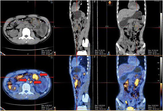
What is Nuclear Medicine?
Nuclear Medicine is a type of advanced imaging in which special technology is used to “watch” certain processes within your body to better understand if and where an issue may be occurring. Radioactive isotopes are injected or ingested by the patient and a set amount of time is allowed for circulation of those isotopes before imaging begins. Your healthcare team is able to get pinpoint answers from this type of process, as the isotopes are demonstrating your body’s processes at a molecular level.
What is Nuclear Medicine for?
Generally speaking, Nuclear Medicine is used for very specific, very specialized medical reasons to study organ or tissue function. While Nuclear Medicine has many different applications, some of the most common uses are for diagnosis of kidney problems, thyroid function, bone scans seeking the cause of pain or inflammation, diagnosis of infectious disease, and locating the cause of heart, brain, or breast issues.
But wait, there’s more!
There are a ton of other radiology procedures, but these are among the most common, and we want to keep this simple. We hope you learned something from this “Radiology for Dummies” guide. If you’re interested in hearing more, check out Episode 21 of Healthcare is Selfcare: The Podcast, with Radiology Manager Jarrod Fetters.
Content Inspired by Healthcare is Selfcare: The Podcast
Radiologists are essentially healthcare photographers. By using specialized technology, or different cameras to take images of the body, they are able to take a closer look at underlying conditions. In this episode of Healthcare is Selfcare: The Podcast, Radiology Manager Jarrod Fetters walks us through the capabilities of imaging.